I. 무기폐(atelectasis) 란?
- 폐실질의 collapse or incomplete expansion, 공기의 감소로 인해 폐의 용적이 줄어든 것
- subsegmental atelectasis + mild fever는 수술후 환자에서 흔하게 볼수 있다.
■ 무기폐(atelectasis)의 원인
1) Obstructive atelectasis
- 폐포 공기가 폐포 모세혈관으로 흡수되었으나, 기관지의 폐쇄로 인해 흡기 공기로 대체되지 않은것
- lobar atelectasis 를 만들고, complete collapse
- supplemental O2를 이용할때 더 발생 (폐포의 흡수속도 산소 > 질소)
- 소아에서는 이물흡인으로 인해 주로 발생하며, 반대편 폐의 과팽창소견이 이때는 나타난다
- ICU 환자에서는 폐포폐쇄로 인해 체액삼출이 동반하여 consolidation과 병합하여 나타날수 있음
2) Relaxation(passive) atelectasis
- 흉강내 mass effect로 인해 발생함
- ex. pleural effusion, pneumothorax, pulmonary mass
3) Adhesive atelectasis
- surfactant(계면활성제) 부족으로 인해 발생함
- neonatal respiratory distress syndrome, ARDS
4) Cicatricial atelectasis
- 폐실질의 섬유화로 인해 용적감소
II. 무기폐(atelectasis)의 영상소견
■ 무기폐의 징후
- displacement of the fissure
- vascular marking crowding
- increased radiopactiy by loss of areation
- elevation of diaphragm
- rib crowding on the side + volume ↓
- Mediastinal shift to the side + volume ↓
- overinflation of adjacent or contralateral lobes
■ Lobar atelectasis 의 소견
- 주로 central bronchial obstruction에 의해 많이 발생함
- lobar atelectasis 환자에서 central tumor은 r/o 되어야함.
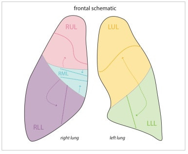
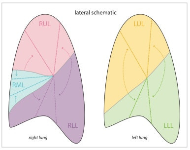
§ Left upper lobe atelectasis
- luftsichel sign(초승달 모양 공기음영)이 관찰된다
- Lateral view 에서 oblique fissure가 anterior 방향으로 이동하거나, wedge-shaped collapse 관찰가능
- 좌엽의 hazy opacity가 관찰됨
§ Left lower lobe atelectasis
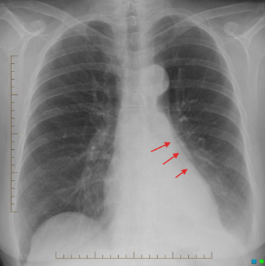
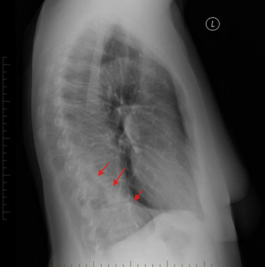
- Triangular retrocardiac opacity ; 좌측심장과 두개의 선으로 보이며, collapse 된 좌하엽이 삼각형으로 보인다
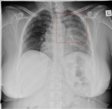
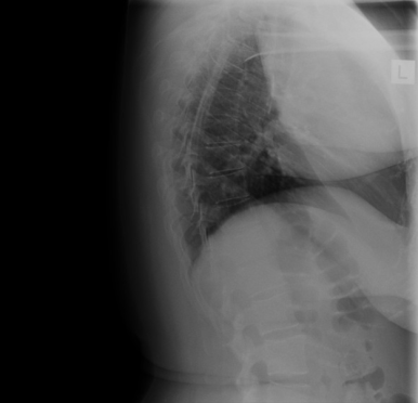
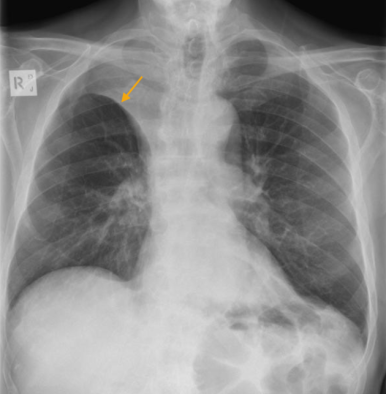
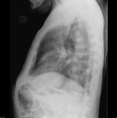
§ Right upper lobe atelectasis
- 우측 폐상엽에서 collapse 된 RUL을 확인 할수 있다
※ Golden S sign
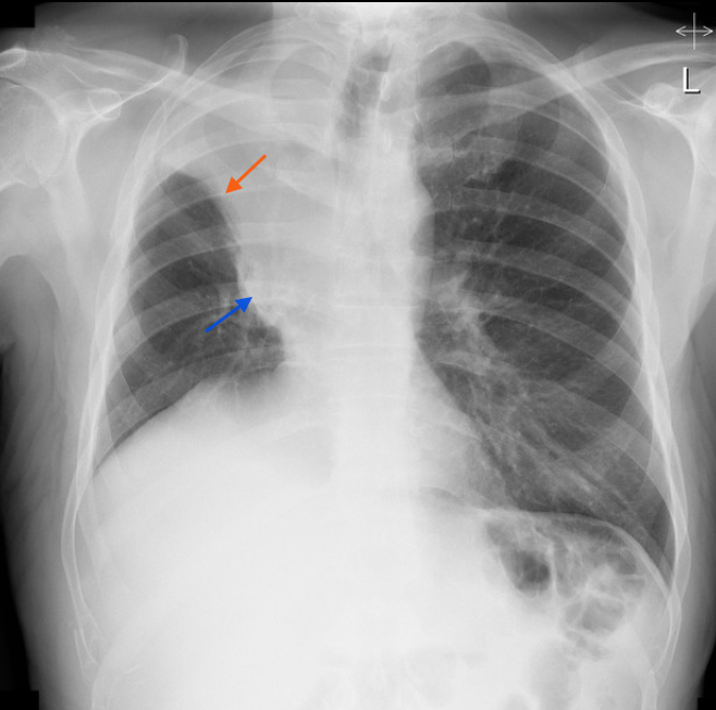
- RUL collapse가 obstructing mass에 의해 발생한 경우 관찰할수 있다.
- 우측 minor(horizontal)fissure displacement (붉은 화살표)
- convex shape mass 가 관찰된다.(푸른 화살표)
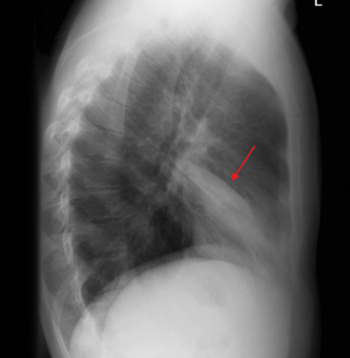
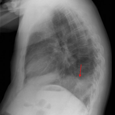
§ Right middle lobe atelectasis
- RML collpase 에서 폐 아래쪽 음영이 약간 증가하고, 실루엣징후가 양성으로 나타날 수 있다.
- lateral view 에서 쐐기 모양 opacity를 확인 할수 있다
§ Right lower lobe atelectasis
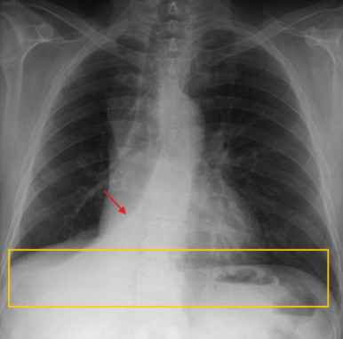
- RLL collapse는 wedge-shaped 이면서 retrocardiac opacity로 나타난다
- 실루엣 징후 양성일수 있다(우측 가로막 경계의 소실)
『출처 : CORE Radiology』
『출처: https://radiopaedia.org/』
'영상의학' 카테고리의 다른 글
| 폐부종(pulmonary edema)의 X-ray, CT 소견 | support devices (0) | 2025.03.24 |
|---|---|
| 공동성 병변, 낭성 병변, 섬유성 변화 | 폐 질환의 주요 영상소견 | CT Xray (1) | 2025.02.23 |
| 흉부 CT 와 X-ray 판독과 이상소견 | 흉부 영상의학 | 이차폐소엽 (0) | 2025.02.17 |