I. 폐 해부학 : Lobes, Segements, Fissure

|
우엽
|
좌엽
|
|
3 lobes, 10 segments
|
2 lobes, 8 segments
|
|
RUL : apical, posterior, anterior
RML : lateral ,medial
RLL : superior, medial basal, anterior basal, lateral basal, posterior basal
|
LUL : apico-posterior, anterior, superior lingular, inferior lingular
LLL : superior, medial basal, anterior basal, lateral basal, posterior basal
|
|
minor fissure (horizontal) : RUL / RML _ 전,측면
major fissure (oblique) : RUL,RML / RLL _측면만
|
major fissure (oblique) : LUL / LLL _ 측면만
|
II. secondary pulomonary nodule (SPL, 이차폐소엽)
■ SPL의 해부학적 구조
- SPL : elemental unit of lung function , 1-2.5 cm 크기 CT에서 보이는 가장 작은 폐 단위
- SPL 마다 centrilobular artery와 bronchus가 존재하고 acinar artery , bronchus로 분지 한다.
- Acinus : 가스교환의 기본유닛, 대략 12 acinus ⊂ SPL
- centrilobular artery : CT에서 희미한 점으로 보임
- centrilobular bronchus : 일반적으로 보이진 않음
- pulmonary vein, lymphatics → SPL 주변 사이사이를 지남, SPL 사이 결합조직 = interlobular septa
■ SPL에서 관찰되는 이상소견
§ Consolidation (경화)
- consolidation 소견
· alveoli가 완전히 액체성 물질 (blood, pus, water, cell) 따위로 채워진 것.
· pulmonary vessels 은 unenhanced CT에서 보이지 않음
· Air bronchogram : consolidation 사이에서 기관지모양 radio-lucent 한 음영이 보이는 것
· Silhouette sign : 같은 plane에서 비슷한 density 물질이 x-ray 상에서 경계가 없어지는 것.
( 심장의 우측경계 : RML, 심장의 좌측경계 : Lt. lingula, 우측가로막 : RLL, 좌측가로막 : LLL )
- Consolidation의 원인
· acute consolidation
> Pnuemonia
> Pulmonary hemorrage
> ARDS
> Pulmonary edema(uncommon)
· chronic consolidation
> Bronchioloalveolar carcinoma
> Organizing pneumonia
> Chronic eosinophilic pneumonia
§ Ground glass opacification (GGO, 간유리음영)
- 소견 cmv 폐렴
· alveoli가 부분적으로 액체성 물질 (blood, pus, water, cell) 따위로 채워진 것
· 폐포벽의 비후 또는 폐포의 공기가 줄어(ex. 무기폐) 발생
· 주로 CT를 통해서 보며, hazy, gauze like opacity를 띄며, 폐혈관은 보임
- GGO의 원인
· acute GGO
> Pulmonary edema(common)
> Pneumonia (atypical type : viral, PCP)
> Pulmonary hemorrhage
> ARDS
· chronic GGO
> Bronchioloalveolar carcinoma : may be focal or multifocal
> Organizing pneumonia : typically rounded and peripheral
> Chronic eosinophilic pneumonia : upper lobe predominace
> Idiopathic pneumonias
> Hypersensitivity pneumonitis : subacute type ; mosaic pattern
> Alveolar proteinosis : typically central to periphery distribution
|
※ Distribution type of GGO
|
|
|
central
|
peripheral
|
|
|
§ Interlobular septal thickening
- smooth interlobular septal thickening 소견
· pulmonary vein dilatation
- smooth interlobular septal thickening 원인 ≒ central GGO ddx.
> Pulmonary edema (m/c)
> Pulmonary alveolar proteniosis
> Pulmonary hemorrhage
> Atypical pneumonia (Pneumocystis jiroveci)
- nodular, irregular interlobular septal thickening 소견
· SPL peripheral lymphatics의 infiltrate에 의해 발생
- nodular, irregular interlobular septal thickening 원인
> Lymphangitic carcinomatosis
> Sarcoidosis
§ Crazy paving pattern
- crazy paving 소견
· interlobular septal thickening + GGO
- crazy paving 원인
> Alveolar proteinosis
> Pneumocystis jiroveci pneumonia
> Organizing pneumonia
> Bronchioloalveolar carcioma
> Lipoid pneumonia (by lipid aspriation)
> ARDS
> Pulmonary hemorrage
■ SPL에서 nodules
§ Centrilobular nodules
- centrilobular nodules 소견

· centrilobular bronchiole(or artery)의 opacification
· SPL 중심의 다발성 결절로 보이며 GGO~solid attenuation
- centrilobular nodules 원인 - 감염성
> Endobronchial spread of TB
> Atypical pneumonia (mycoplasma)
> Bronchopneumonia
- centrilobular nodules 원인 - 염증성
> Hypersensitivity pneumonitis (HSP)
> Respriatory bronchiolitis intersitital lung disease (RB-ILD)
> Silicosis
> Diffuse panbronchiolitis
§ Perilymphatic nodules
- perilymphatic nodules 소견


· subpleural / pribronchovascular / septal → 위치에 따라 나눌 수 있음
- perilymphatic nodules 원인
> Sarcoidosis
> Pneumoconioses (진폐증)
> Lymphangitic carcinomatosis
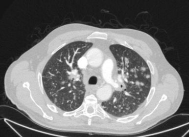
§ Randomly distributed nodules - 혈행성전파
- randomly distributed nodules 원인


> Hematogenous metastaes
> Septic emboli
> Pulmonary Langerhans's cell histocytosis (PLCH)
§ military nodules - 혈행성전파
- military nodules 원인

> Disseminated TB
> Disseminated Fungal infection
> Disseminated hematogenous metastases
§ Tree-in-bud nodules


- Tree-in-bud nodules 원인
> M.tuberculosis, atypical mycobacteria
> Invasive aspergillus
『출처 : CORE Radiology』
『출처: https://radiopaedia.org/』
'영상판독' 카테고리의 다른 글
| 폐부종(pulmonary edema)의 X-ray, CT 소견 | support devices (0) | 2025.03.24 |
|---|---|
| 공동성 병변, 낭성 병변, 섬유성 변화 | 폐 질환의 주요 영상소견 | CT Xray (1) | 2025.02.23 |
| 무기폐의 진단과 x-ray 소견 | 무기폐의 원인과 위치 | atelectasis (0) | 2025.02.20 |