반응형

영상의학 4

폐부종(pulmonary edema)의 X-ray, CT 소견 | support devices

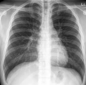
♠ Pulmonary Edema (폐부종)§ 폐부종의 임상적 특징 - acute breathlessness - orthopnea - paroxysmal nocturnal dyspnea - foaming at the mouth - distress§ 폐부종의 영상학적인 중증도는 3단계로 나눌 수 있고, pulmonary venous pressures에 따라 결정된다. grade 0: normal chest radiograph, PCWP 8-12 mmHggrade 1: upper lobe diversion on a chest radiograph, PCWP 13-18 mmHggrade 2: interstitial edema on a chest radiograph, PCWP 19-25 mmHggrade 3: al..

영상의학 2025.03.24

공동성 병변, 낭성 병변, 섬유성 변화 | 폐 질환의 주요 영상소견 | CT Xray

■ Cavitary lesion / nodule (공동성 병변) § Solitary cavitary nodules (단일 공동성 결절) - 공동성 병변은 thick, irregular 한 wall- 내부에 solid mural component가 존재할 수도 있음. - 벽의 두께에 따라 4mm 이하 = benign, 15mm 이상일 경우 malignant의 가능성이 높음 - Spiculated(침상형)의 경계를 가질경우 malignant의 가능성 높음    - Primary bronchogenic carcinoma→ ADC와 SCC 모두 공동을 형성 할수 있으나, SCC에서 더 흔함. SCLC는 공동 형성 X - Tuberculosis → 고전적으로 폐상부에 위치하는 공동성 병변은 결핵을 시사함.  §..

영상의학 2025.02.23

무기폐의 진단과 x-ray 소견 | 무기폐의 원인과 위치 | atelectasis

I. 무기폐(atelectasis) 란? - 폐실질의 collapse or incomplete expansion, 공기의 감소로 인해 폐의 용적이 줄어든 것 - subsegmental atelectasis + mild fever는 수술후 환자에서 흔하게 볼수 있다.​■ 무기폐(atelectasis)의 원인 1) Obstructive atelectasis - 폐포 공기가 폐포 모세혈관으로 흡수되었으나, 기관지의 폐쇄로 인해 흡기 공기로 대체되지 않은것 - lobar atelectasis 를 만들고, complete collapse - supplemental O2를 이용할때 더 발생 (폐포의 흡수속도 산소 > 질소) - 소아에서는 이물흡인으로 인해 주로 발생하며, 반대편 폐의 과팽창소견이 이때는 나타난다..

영상의학 2025.02.20

흉부 CT 와 X-ray 판독과 이상소견 | 흉부 영상의학 | 이차폐소엽

I. 폐 해부학 : Lobes, Segements, Fissure우엽좌엽3 lobes, 10 segments​​2 lobes, 8 segmentsRUL : apical, posterior, anteriorRML : lateral ,medialRLL : superior, medial basal, anterior basal, lateral basal, posterior basalLUL : apico-posterior, anterior, superior lingular, inferior lingularLLL : superior, medial basal, anterior basal, lateral basal, posterior basalminor fissure (horizontal) : RUL / RML _ 전..

영상의학 2025.02.17
반응형