I. 폐암의 영상학적 소견
§ solitary pulmonary nodule or lung mass
- Adenocarcinoma의 50%는 solitary pulmonary nodule로 나타남
§ Segmental or lobar atelectasis
- 기관지 폐쇄로 인한 무기폐는 흔한 폐암의 presentation
- 폐쇄성 폐렴은 기관지폐쇄와 폐실질의 경화소견이 나타남
- 두 개 이상의 무기폐 소견은 양성일 가능성이 높지만, CT와 기관지 내시경을 통한 검사는 필요함
§ Consolidation
- 폐렴과 BAC(mucinous subtype)에서 나타난 경화 소견은 구분하기 어려움
- 해소되지 않는 경화소견, 정상 wbc count가 특징
§ Hilar mass
- small cell carcinoma와 squamous cell carcinoma의 흔한 presentation
- hilar enlargement는 primary central tumor 또는 폐실질 neoplasm의 전이
- Tumor가 bronchus를 압박하는 경우 가 많다. 좁아진 기관지는 폐암에 특이적 소견
§ Superior sulcus tumor
- lung apex에 발생한 폐암
- Pancost tumor : sympathetic ganglia를 침범하여 Honer syndrome을 발생 (ipsilateral ptosis, anhidrosis)
§ Lymphangitic carcinomatosis
- diffuse spread of neoplasm typically seen in late-stage disease
-주로 비대칭적 nodular interlobular septal thickening 소견
§ Pleural effusion
- pleural metatasis or lymphatic obstruction으로 인해 발생함
§ Pneumothorax
- peripheral tumor가 흉막으로 침범하거나 공동을 형성하여 발생함
♠ 폐암의 영상소견 자세히 알아보기▶ https://medhamstern.tistory.com/165
II. Staging of lung cancer (폐암의 병기)
■ Stage groups of TMN stage

■ T stage
● T1
- 3cm 이하 tumor면서 lung or visceral plerua로 둘러싸여 있음.
- T1a : 1cm 이하, T1b : 2cm 이하, T1c : 3cm 이하
● T2
- 3cm 초과 이면서 5cm 이하 tumor
- visceral pleura의 침범
- main bronchus를 침범했으나, carina involvement X
- T2a : 3cm 초과 4cm 이하. T2b : 4cm 초과 5cm 이하
● T3

- 5cm 초과 7cm 이하 tumor
- Phrenic nerve, Parietal pericardium, Azygos vein 침범
- Parietal parietal pleura, thoracic T1 T2 nerve root, stellate ganglion 침범
- Separate tumor nodules in the lobe of the primary (동측, 같은엽내 존재)
● T4
- 7cm 초과 tumor
- Carina를 침범
- Trachea, 대혈관(ex.SVC) 침범
- diaphragm 침범
- Vertebral body 침범
- Mediastinum 침범 (가슴샘, 심장, 미주신경, 식도 등등)
- Tumor accompanied by ipsilateral separate tumor nodules, different lobe
(동측 폐 다른 엽에 nodules 존재)
- subcalvian vessels, spinal canal cervical nerve root or brachial plexus 침범
■ N stage
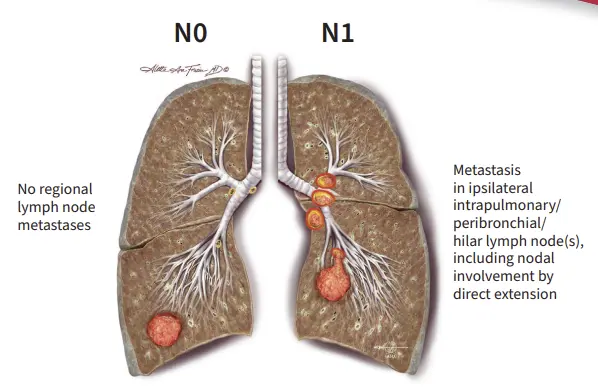
● N0
- No lymph node metastases
● N1
- Ipsilateral hilar or Intrapulmonary lymph node(같은 쪽 폐 내부 or 기관지 림프절)
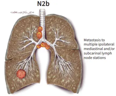
● N2
- N2a : Metastasis to single ipsilateral mediastinal or subcarinal LN (같은쪽 종격동 림프절)
- N2b : Metastasis to multiple ipsilateral mediastinal or subcarinal LN

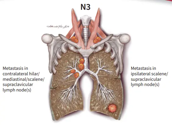
● N3
- Metastasis in contralateral hilar/mediastinal/scalene/supraclavicular LN (반대편 림프절)
- Metastasis in ipsilateral scalene/supraclavicular LN (양측 쇄골상부 림프절)
■ M stage
● M0
- No metastasis
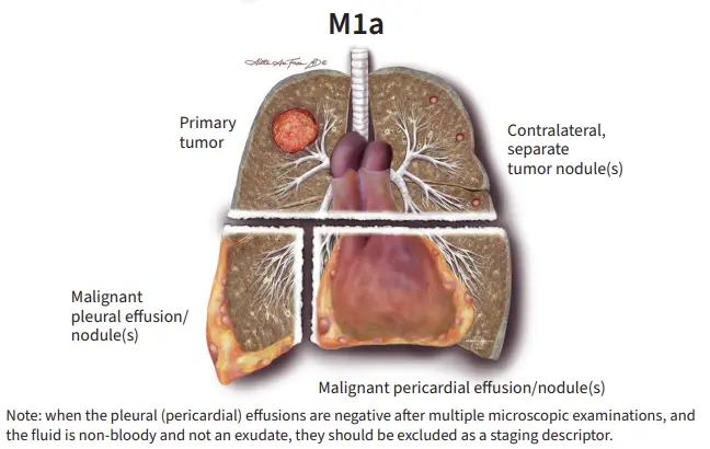
● M1a
- Contralateral separate tumor nodules (반대편 다른 엽에 nodule)
- Malignant Pleural effusion
- Malignant Pericardial effusion (삼출액의 발생)
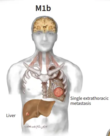
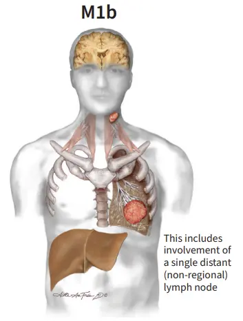
● M1b
- single distant or extrathoracic metastasis
● M1c
- M1c1 : Multiple extrathoracic metastases in single organ system
- M1c2 : Multiple extrathoracic metastases in multiple organ system

『출처 : CORE Radiology』
『출처: https://radiopaedia.org/』
『출처 :Staging Cards in Thoracic Oncology, 9th Edition, IASLC 』
'호흡기' 카테고리의 다른 글
| 천식의 치료단계 조절 | 천식의 비약물치료 | 진료치침 (0) | 2025.03.28 |
|---|---|
| 천식의 원인과 종류 | 천식의 진단 기준과 폐기능검사와 기관지유발검사 (0) | 2025.03.27 |
| 고립성폐결절 (Solitary pulmonary nodule)의 영상소견 CT 검사 | 폐암의 조직학적 분류 | 영상소견 (1) (0) | 2025.03.25 |
| 아스페르길루스 감염증 | 진균종 aspergilloma | aspergillus | 영상소견 | 임상양상 (1) | 2025.03.12 |
| 결핵의 임상양상과 영상소견 | 원발성 결핵, 재활성 결핵, 비결핵 항산균 (0) | 2025.03.06 |