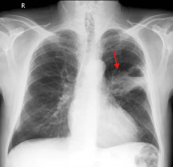
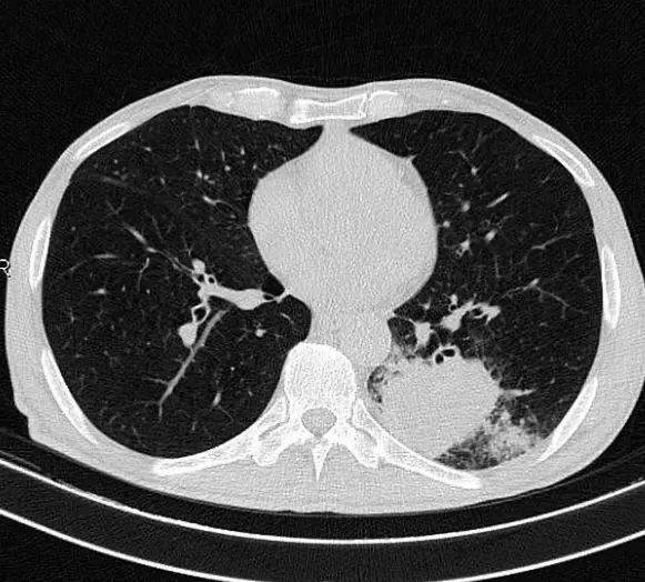
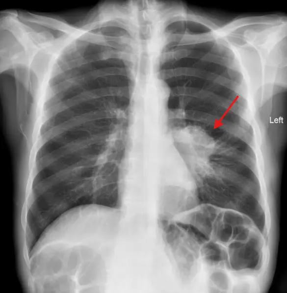
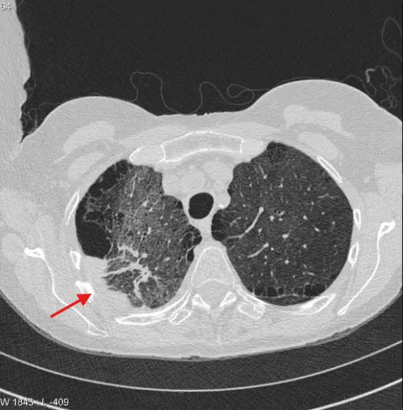
I. Solitary Pulmonary nodule (SPN, 고립성폐결절)
■ SPN 이란?
- 경계가 명확한 원형, 또는 난원형의 3cm 이하 결절
- 3cm 이상일 경우 종괴(mass)
■ 거의 확실하게 양성을 시사하는 SPN
- Central, Laminar, Diffuse calcification
- Popcorn calcification (pulmonary harmartoma)
- Intra-lesional fat (harmartoma or lipoid granuloma)

■ 양성을 시사하는 SPN
- small nodules (3mm 미만 = 0.2 % 악성 , 4-7mm = 2.7% 악성)
- any calcificatioin a small nodule
- Non round shape(타원, 다각형, 삼각형...)
- subpleural location
- clustering of nodules (감염 시사)
■ 악성을 시사하는 SPN
- Large size(가장 중요한 인자) (0.8-3cm = 18% 악성, 3cm 초과 시 높은 악성가능성)
- Irregular edge or Spiculated margin(침상 변연)
- 원형모양(타원형, 다각형대비)
- 공동성 결절, 낭성 공간을 포함한 결절
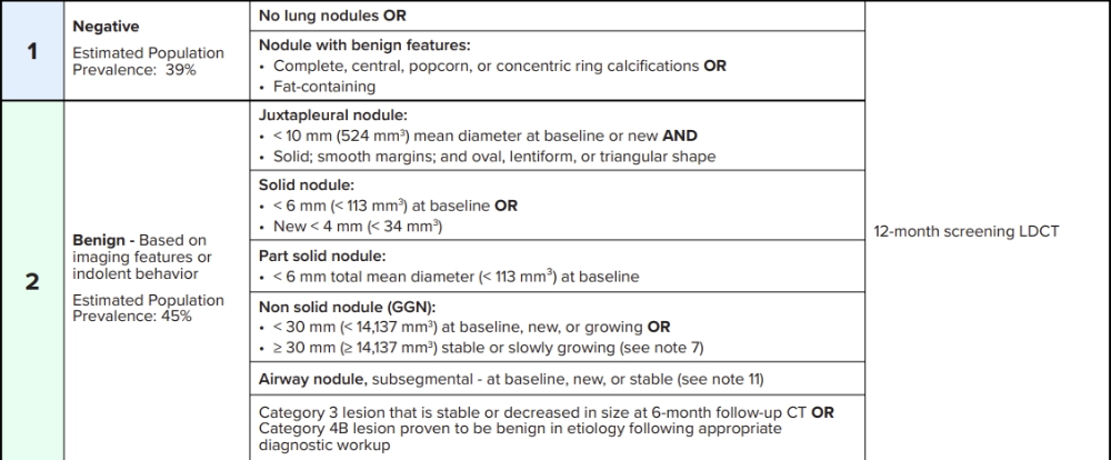
II. SPN f/u, 고립성 폐결절의 검사 (Fleischner Society , 2017)
§ Solid nodule
- 6mm 미만 고형결절
→ low risk patients : no routine follow-up required
→ high risk patients : optional CT at 12 months
- 6-8mm 고형결절
→ CT at 6-12 months then CT 18-24 months
- 8mm 초과 고형 결절
→ CT at 3 months, PET-CT or tissue sampling
★ Lung-RADS ⓒ 2022 American College of Radiology®


III. 페암의 조직학적 분류와 영상소견 정리
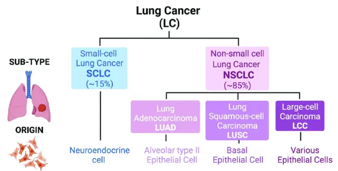
§ Adenocarcinoma
- 가장 흔한 폐암의 subtype, 흡연과 관련성이 있으나 Squamous cell caricinoma 대비 연관성은 낮음
- Peripheral lung에 발생하는 경향
- 전형적 소견 : Pulmonary nodule with spiculated margin by reactive fibrosis
- Cavitation 도 발생가능하지만, Squamous cell 보단 흔하지 않음
- TTF-1 (thyroid transcription factor) : primary lung adenocarcinoma marker
§ Bronchioloalveolar carcinoma (BAC)
- 폐실질의 침윤과 파괴를 통한 성장(hilic growth)을 하는 다른 폐암과 달리 폐포벽을 따라 성장하는 well differentiated adenocarcinoma (lepidic growth)
- 완만한 진행을 보이는 경우가 많고 PET 음성인 경우가 많다
- 100% 생존율 작은 종양 ~ 광범위 진행까지 다양한 스펙트럼
▶ BAC의 분류
- Adenomatous hyperplasia
- Adenocarcinoma in situ
- Minimally invasive adenocarcinoma
- non mucinous BAC
- mucinous BAC(Invasive mucinous carcinoma)
● Non mucinous BAC (lepidic predominant ADC)
- 영상 : ground glass or solid nodule with air bronchograms
- mucinous subtype 보다 좋은 예후
● Mucinous BAC
- 영상 : chronic consolidation


§ Squamous cell carcinoma
- 과거 가장 폐암의 흔한 subtype
- Main, lobar, segmental bronchi에서 많이 발생함
- hilar mass 형태로 많이 발견, bronchial obstruction 증상으로 발견됨
- 소견 : Lobar atelectasis, Mucoid impaction, consolidation, bronchiectasis
- 소견: Cavitation 흔함
§ Small cell carcinoma
- 세번째로 흔한 폐암 subtype
- Neuroendocrine origin으로 다양한 부종양증후군과 관련되어 있음
- 흡연과 강한 연관성
- 주로 중심기관지에서 발생 후 기관지벽을 침범, larger hilar or perahilar mass 로 잘 나타남
- SVC 침범 시 SVC syndrome 유발가능
- SPN(단일 폐결절)로 나타나는 경우는 드물다
- 수술은 일반적으로 적합하지 않은 유형

§ Large cell carcinoma
- lung periphery에서 잘 발생하며 큰 종괴로 나타남
- 흡연과 강한 연관성, 예후 안 좋음
§ Carcinoid tumor
- 기관지 벽의 neuroendocrine cell 기원
- carina 원위부 기관지 내 종괴로 나타남, 폐쇄성 무기폐를 유발할 수 있음
- 20% 정도는 SPN으로 나타남
- Typical(low-grade)과 Atypical(aggressive) 타입으로 분류됨.
- 림프절 전이 및 원격전이가 없는 typical carcinoid는 예후 좋음 (5년 생존율 92%)
- DIPNECH : carcinoid의 드문 전구병변, 작은 종양과 폐쇄성 세기관지염


『출처 : CORE Radiology』
『출처: https://radiopaedia.org/』
'호흡기' 카테고리의 다른 글
| 천식의 원인과 종류 | 천식의 진단 기준과 폐기능검사와 기관지유발검사 (0) | 2025.03.27 |
|---|---|
| 폐암 TNM 분류를 통한 병기 설정 | 크기, 침범부위, 전이 여부에 따른 lung cancer staging (0) | 2025.03.26 |
| 아스페르길루스 감염증 | 진균종 aspergilloma | aspergillus | 영상소견 | 임상양상 (1) | 2025.03.12 |
| 결핵의 임상양상과 영상소견 | 원발성 결핵, 재활성 결핵, 비결핵 항산균 (0) | 2025.03.06 |
| 폐렴의 임상적 분류와 영상학적 패턴 | 폐렴의 합병증 영상소견 (0) | 2025.02.28 |